
How Can We Help?
Paraparesis
Paraparesis
There are only a limited number of neurological emergencies, and it is often forgotten that sudden onset of weakness in both legs is one of them. Consider the following:
- Spinal tuberculosis.
- Malignancy.
- Guillain-Barre syndrome.
- HIV myelopathy
- Spinal cord infarction (aortic disease, syphilis)
- Vitamin B12 deficiency
- Trauma.
The key to management is recognition that the history is often confusing, and what is presented as an acute event may have been progressing over days to weeks. Early vigorous investigation is usually warranted, and if in any doubt about the relative urgency, ask for advice.
Lower motor neurone weakness with sphincter sparing and absent (or rapidly disappearing) reflexes– acute polyneuropathy (Guillain-Barre). Monitor respiration and consider ventilation if FVC less than 1.5 litres. (Note that you cannot use a peak flow meter to monitor lung volumes, and the use of accessory muscles as a visible sign of respiratory distress, familiar from other conditions, may be less obvious if the muscles themselves are weak.)
Wasting/clubbing/hard cervical adenopathy/mass on CXR. Most likely to be a malignancy with vertebral spread.
Upgoing plantars with absent ankle jerks – consider B12 deficiency. Folate supplementation as a precipitant of acute deterioration in B12 deficiency has long been considered an important aetiology, but may well have been based on unreliable observational data, and in any case doesn’t make biochemical sense.1
Spinal tenderness, no obvious features to suggest malignancy – consider spinal tuberculosis. Sometimes the double shadow of the spinal abscess is clearly visible behind the heart on a plain CXR.
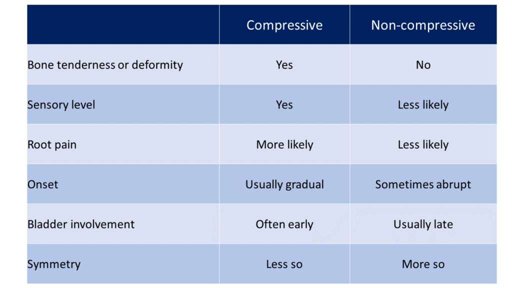
Paraparesis management:
Emergency radiotherapy plus dexamethasone in high doses – e.g. 8 mg 8 hourly – may help patients with malignancy.
Surgical decompression of TB abscesses gives good results if done timeously.
If there is any possibility of B12 deficiency, take blood for serum B12, then treat immediately with IM Vitamin B12 1 mg daily for a week while awaiting results.
Patients with suspected Guillain-Barre should be managed in a location where respiration can be closely monitored and ventilation is feasible – in some series as many as 25% required some form of ventilatory support.
Perspective – spinal tuberculosis.
Rare variants of tuberculosis causing neurological deficit have been described, such as patients with tuberculomas of the cord2 and patients with hemiplegias or monoplegias presumed to be because of vascular involvement at a dens level leading to a medial medullary syndrome.3
There is a dearth of good quality information on management issues in this field. The MRC Working Party on tuberculosis of the spine has published a whole series of reports. One4 suggests rather unsurprisingly that 6-9 months of therapy with a rifampicin-containing regimen is as good as longer courses of therapy with poorer quality regimens, however these trials have been criticised because of issues with sample size and statistical analysis.
A RCT from India5 demonstrated that in patients without neurological deficit, standard nine month chemotherapy without surgery resulted in 99% of patients being well at ten year follow-up; no surgery with only six months of TB treatment resulted in 94% of patients being well at ten years. Surgery plus six months of treatment yielded the worst outcome (90% well at ten years.)
The authors of larger retrospective management series6,7 are in favour of prompt surgical decompression if there is any neurological deficit, although the quality of evidence is not good.
Cord compression
Extrinsic compression of the cord can be due to a vast array of pathologies, although disease of the vertebral bodies is probably the commonest (tuberculosis, metastatic malignancy or trauma). Rarities such as hydatids and cryptococcomas are described, and can sometimes be suspected pre-operatively. The site of compression of the cord may not be exactly that suggested by the sensory level8– it is often higher (e.g. a sensory level at T8 may turn out to be due to a lesion at T4). This is thought to be due to the somewhat inconsistent vascular supply of the cord.
Work up of cord compression:
- History – onset, duration of weakness, features suggestive of systemic illness.
- Examination – sensory level, tone, sphincters, grade power in ankles and hips.
- Spinal tenderness or deformity?
- Significant adenopathy – specifically check for a VT node.
- X-ray the thoracic spine and do a CXR. CT or MRI is often helpful (discuss.)
- If the CXR is highly suggestive of malignancy or multiple metastases are seen radiologically, then aim for a tissue or cytological diagnosis.
- If there is vertebral collapse and nothing else to suggest malignancy, try to get a decompression/biopsy done.
- If clearly malignant try to organise radiotherapy.
Guillain-Barre syndrome
Presentation
- Progressive weakness of more than one limb
- Duration less than 4 weeks
- Lower motor neurone type (flaccidity and reduced reflexes)
- No other obvious cause
- Urinary symptoms can be present in about a quarter of patients, with retention in nearly 10%9
There are several variants:
- Acute inflammatory demyelinating polyneuropathy (AIDP) accounts for about 75% of patients. Predominantly motor, but mild sensory changes.
- Acute motor axonal neuropathy (AMAN).
- Acute motor and sensory axonal neuropathy (AMSAN) is rare.
- Miller Fischer syndrome – ophthalmoplegia, ataxia, areflexia, but not weak.
Management of Guillain-Barre
- Mortality should be less than 5% if patients are ventilated if necessary[38].
- Monitor FVC and ventilate if needed (FVC less than 1.5 litres or falling fast, or <20ml/kg, or > 30% drop from baseline)
- Heparin for DVT prophylaxis
- Adequate nutrition.
- Corticosteroids don’t help and should not be given (but see under HIV).
- Plasma exchange does work but is technically challenging and very expensive.
- Intravenous immunoglobulin is also very expensive but works as well as plasma exchange
Perspective – plasma exchange and immunoglobulins in Guillain-Barre.
There is reasonable evidence that plasma exchange is of benefit, and there is some evidence to suggest that intravenous immunoglobulin achieves the same benefits as plasma exchange. There are no trials of adequate quality comparing immunoglobulin to placebo, but it is usually accepted that it is superior in view of the probable equivalence to plasma exchange. The size of the benefit is worth reviewing[39]:
Outcome measure | Plasma exchange vs. placebo | 95% Confidence interval | Immunoglobulin vs. plasma exchange | 95% Confidence interval |
|---|---|---|---|---|
Mean days of ventilation | – 5 | -13 to +3 | Median about 30 days both groups | No significant difference |
Mortality at 1 year (ARR) | 0.8% | -2.7% to+ 4.3% | 0.2% | -3.3% to +3.8% |
Walk with aid after 4/52(ARR) | 0.1% | -8% to +8% | Inadequate data | |
Walk without aid at 4/52 (ARR) | 7.8% | 0% to 15.5% | Median time 50 to 60 days both groups | No significant difference |
Full strength at 1 year | 13.2% | 3.7% to 22.4% | Inadequate data |
The systematic review failed to demonstrate a statistically significant difference in mortality or ventilation time but showed an absolute risk reduction for walking without aid at one month of 8%. (NNT =13.) IVIG and PE appear similar in efficacy[40]. The clinical relevance of these differences in a condition with a fairly arduous rehabilitation process lasting about two months involves a number of value judgements, one of which might be the safety and cost of ventilation in an individual institution.
Timing. The reviews cited above suggested that benefit was higher the earlier treatment was given (best before 7 days) but there was some evidence of benefit even up to 30 days.
Transverse myelitis
Terminology: An inflammatory process (of any cause!) involving only the grey matter is called poliomyelitis; if it is confined to the white matter, it is called leukomyelitis; if it involves both it is called a ‘transverse myelitis’
Causes:
- Viruses – herpes (dorsal root ganglion), HIV and polio (anterior horn)
- Bacteria – tuberculosis or syphilis
- Parasitic – hydatid or cysticercosis
- Immune-mediated – multiple sclerosis, post-vaccine.
A separate but important entity is a spinal epidural abscess, which may be due to tuberculosis or to pyogenic organisms, particularly staph:
- Fever and headache
- Localised back pain
- Then neck stiffness and progressive paraplegia.
If not thought about carefully first, such individuals are often subjected to lumbar puncture, which can be rather confusing – some cells, usually less than 100, and not always all polymorphs, a raised protein, and a normal glucose. (i.e. classical neighbourhood syndrome CSF findings). The clinical picture due to tuberculosis is very similar although with a more indolent onset. (Look for a double shadow on thoracic or plain CXR, often clearly visible ‘behind’ the heart, which may signify extravertebral extension.) Management of both is primarily surgical, and urgently so.
Vascular causes of cord pathology.
A similar syndrome that is not strictly myelitis is due to vascular lesions of the cord. The cord has a tenuous and rather variable blood supply, and infarcts of the cord can occur either due to degenerative vascular disease or vasculitis (e.g. SLE, syphilis). The characteristic feature of infarction is bilateral (usually) motor function loss in the legs which is often incomplete and accompanied by sensory loss which spares the posterior columns (they are supplied by the posterior spinal artery and not by the anterior median spinal artery which supplies most of the rest of the cord). The sensory loss is thus for pain and temperature but not vibration or position sense. Very occasionally, the infarct can be confined to the grey matter anteriorly, in which case there is paraplegic upper motor neurone type motor weakness without any sensory involvement.
Perspective – Elsberg phenomenon
In cervical spinal extradural compression, one presentation is with ipsilateral arm weakness followed by weakness of the leg on the same side, then the opposite leg, and finally the contralateral arm – so-called U-shaped/clockwise progression or Elsberg phenomenon. Proposed mechanisms are either arterial or direct compressive/venous.
Arterial supply mechanism: The lateral corticospinal tract has long end artery from anterior spinal artery supplying the arm, and arterial vasocorona supplying the dorsal part with the more posterior (leg) part of the LCS. So an extramedullary intradural mass would first involve the long end artery from ASA, then the vasocorona on the same side, then because of the cord being pushed to the other side, vasocorona on contra lateral side to involve that leg, and finally the relatively spared anterior spinal vessel to affect contralateral arm. An alternative more tenuous but still plausible explanation involves a similar sort of process involving venous drainage and leading to venous infarction/thrombosis.
Spinal cord injury without radiological abnormalities (SCIWORA)
This is another relatively rare cord process where a traumatic neck or back injury is followed by either quadriplegia or paraplegia without any clear radiological changes on CT scan, although cord oedema may be identified on MRI10. Management is expectant, although some authorities recommend a course or corticosteroids, and this is quite tempting in the face of devastating neurolgical deficits.
Although quite murky, the pathophysiology may be related to that of a rather similar named condition – SPAM (Subacute Posttraumatic Ascending Myelopathy) where an initial neurological deficit following a cord injury then progresses over the next few days.11
Berry RJ. Lack of historical evidence to support folic acid exacerbation of the neuropathy caused by vitamin B12 deficiency. Am J Clin Nutr. 2019 Sep 1;110(3):554-561. doi: 10.1093/ajcn/nqz089. PMID: 31187858; PMCID: PMC6785032. ↩
Sharma MC, Arora R, Deol PS, et al. Intramedullary tuberculoma of the spinal cord: a series of 10 cases. Clin Neurol Neurosurg. 2002;104:279-84 ↩
Dhammi IK, Singh S, Jain AK. Hemiplegic/monoplegic presentation of cervical spine (C1-C2) tuberculosis. Eur Spine J. 2001;10:540-4 ↩
MRC Working Party on Tuberculosis of the Spine. Five-year assessment of controlled trials of short-course chemotherapy regimens of 6, 9 or 18 months’ duration for spinal tuberculosis in patients ambulatory from the start or undergoing radical surgery. Fourteenth report of the Medical Research Council Working Party on Tuberculosis of the Spine. Int Orthop. 1999;23:73-81 ↩
Parthasarathy R, Sriram K, Santha T, et al. Short-course chemotherapy for tuberculosis of the spine. A comparison between ambulant treatment and radical surgery – ten-year report. J Bone Joint Surg Br. 1999;81:464-71 ↩
Turgut M. Spinal tuberculosis (Pott’s disease): its clinical presentation, surgical management, and outcome. A survey study on 694 patients. Neurosurg Rev. 2001;24:8-13 ↩
Adendorff JJ, Boeke EJ, Lazarus C. Pott’s paraplegia. S Afr Med J. 1987;71:427-8 ↩
Jamieson DR, Teasdale E, Willison HJ. False localising signs in the spinal cord. BMJ. 1996;312:243-44 ↩
Sakakibara R, Uchiyama T, Kuwabara S, Mori M, Ito T, Yamamoto T, Awa Y, Yamaguchi C, Yuki N, Vernino S, Kishi M, Shirai K. Prevalence and mechanism of bladder dysfunction in Guillain-Barré Syndrome. Neurourol Urodyn. 2009;28(5):432-7. doi: 10.1002/nau.20663. PMID: 19260087. ↩
Atesok K, Tanaka N, O’Brien A, Robinson Y, Pang D, Deinlein D, Manoharan SR, Pittman J, Theiss S. Posttraumatic Spinal Cord Injury without Radiographic Abnormality. Adv Orthop. 2018 Jan 4;2018:7060654. doi: 10.1155/2018/7060654. PMID: 29535875; PMCID: PMC5817293. ↩
Basu, Saumyajit MS(Orth), DNB(Orth), FRCS(Edin)1; Gohil, Kushal MS(Orth), DNB(Orth), FASSIa. Subacute Posttraumatic Ascending Myelopathy After Thoracolumbar Spinal Cord Injury. JBJS Reviews 10(10):e22.00097, October 2022. | DOI: 10.2106/JBJS.RVW.22.00097 ↩